

A variation on traditional CT scanning recently gave archaeologists a much closer look at a 2,400-year-old mummified hand from ancient Egypt. Medical imaging already uses the technology for higher-resolution views of patients' soft tissues, but this is the first time it has been tested on mummified human remains.
For soft tissue imaging, contrast is key
“There has long been a struggle to retrieve information from ancient soft tissues, and neither conventional X-ray imaging and CT, nor new methods like MRI and terahertz imaging, have been able to give images with enough contrast or resolution for detailed analysis of these tissues,” physicist Jenny Romell of the KTH Royal Institute of Technology in Sweden told Ars Technica.
CT scanning relies on X-rays but uses a computer to combine images from different angles into cross-sectional views of the body. Differences in the amounts of radiation absorbed by different materials produce the contrast visible in the resulting image. The method works especially well for hard, dense materials like bone, but most types of soft tissue dont produce enough contrast for really high-resolution imagery. Instead, paleopathologists who want to study ancient tissues have to take small samples from mummified remains and examine them under a microscope.
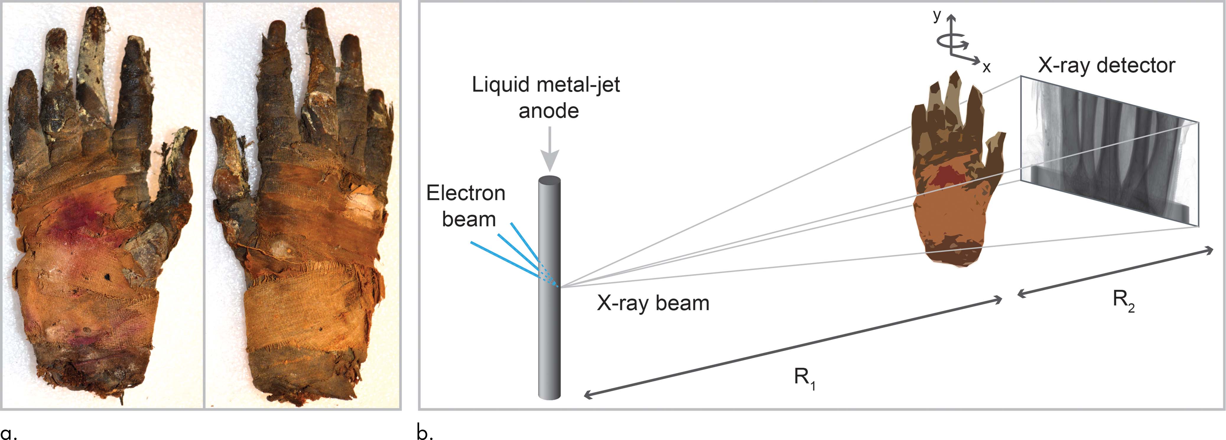
But theres another type of X-ray imaging, called “phase contrast imaging,” that produces higher-resolution images of soft tissue. This method works by measuring something called phase shift, which is a change in the position of a light wave. Romell and her colleagues compare it to the way a ray of visible light gets refracted on its way through a piece of glass. The difference in phase shifts that occur as X-rays pass through different tissues produces much greater contrast, which provides higher-resolution images.
Romell and her colleagues recently tried the method on a mummified hand from ancient Egypt.
A closer look
“Phase-contrast imaging is already used in (for example) biomedical imaging and material science, but this is the first time it has been used to study a mummy,” Romell told Ars Technica. The project began as a chance meeting at a conference, when biomedical physicist Hans Hertz and Egyptologist Salima Ikram started a conversation about imaging mummified remains. They wanted to see how well phase-contrast CT would work on a mummy, and fortunately there was one… well, handy.
The mummified right hand resides at the Museum of Mediterranean and Near Eastern Antiquities in Stockholm, Sweden. It arrived in the late 1800s, along with a skull, two feet, and a handful of other mummified body parts, courtesy of Swedish Count Carlo Lundberg, who had joined the flood of Europeans looting antiquities from sites across Egypt and bringing them home to museums. (Their lack of proper documentation continues to provide modern researchers with occasional headaches.) Its thought to have come from somewhere around Thebes and has been radiocarbon dated to around 400 BCE.
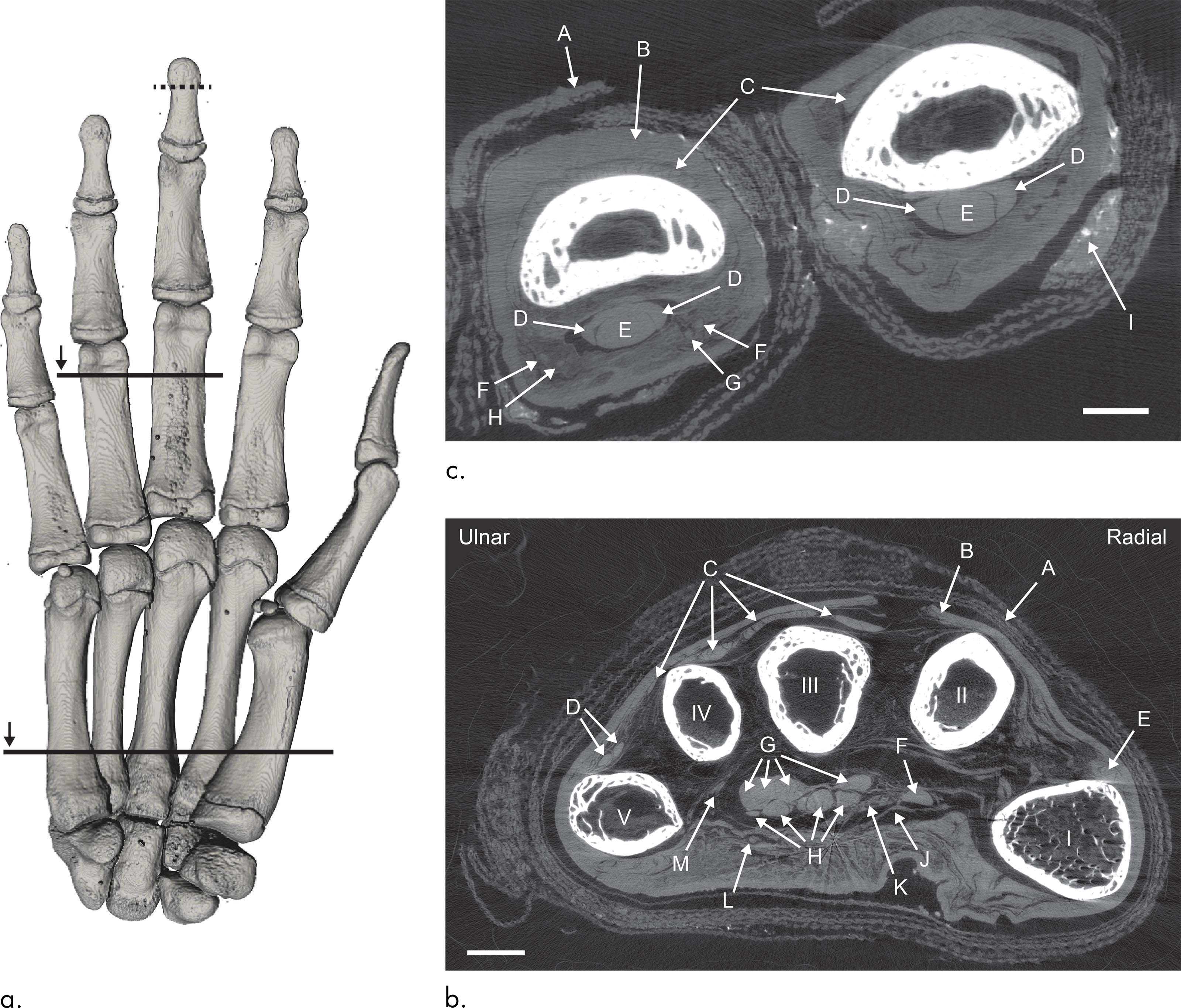
Romell and her colleagues imaged the whole hand, then zoomed in for a closer scan of the tip of the middle finger at a resolution of six to nine micrometers—about the width of a human red blood cell. The results demonstrate that phase-contrast imaging works on mummies—and that the embalmers of Late Period Egypt were very good at their work. Looking at the images, the team could see nerves and the tiny blood vessels in the nail bed. They could recognize different layers of skin, examine the microscopic anatomy of the mummys bone marrow, and even pick out round cells called adipocytes, which store fat.
Whats next?
“Without speculating on exactly what new insights this new technique will bring, we can say that it opens up for non-destructive imaging down to a microscopic level in a way that has not been possible before,” Romell told Ars Technica. She and her colleagues dont have any more mummy experiments planned, but they hope archaeologists and paleopathologists will be able to use phase-contrast CT in place of tissue sampling to study ancient diseases and health.
Of course, knowing that the method works is one thing, and getting access to the equipment is another. Very few archaeology departments have their own CT scanner, so researchers studying mummified remains often have to negotiate with hospitals or medical research facilities for access to CT scanners, MRI machines, and other imaging equipment. Thats likely to be the case for phase-contrast CT, too, although Romell told Ars Technica that the total cost of the equipment is no more expensive than a standard CT scanner.
Radiology, 2018. DOI: 10.1148/radiol.2018180945 (About DOIs).
[contf] [contfnew] 
Ars Technica
[contfnewc] [contfnewc]


